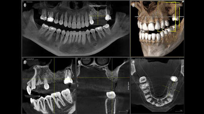
In recent years, the field of dentistry has witnessed remarkable advancements in diagnostic technology, with 3D cone beam computed tomography (CBCT) emerging as a pivotal tool. This cutting-edge imaging technology offers unparalleled detail and precision, allowing dental professionals to enhance their diagnostic capabilities significantly. With 3D cone X-ray imaging, practitioners can visualize the oral and maxillofacial regions in three dimensions, providing comprehensive insights that are essential for accurate diagnosis and treatment planning.
The Advantages of 3D Cone X-Ray Imaging
3D cone beam technology brings forth several advantages over traditional 2D imaging methods. These improvements are crucial for both practitioners and patients, offering a more holistic view and facilitating better clinical outcomes.
- Enhanced detail and accuracy: 3D imaging provides a comprehensive view of the teeth, soft tissues, nerve pathways, and bone in a single scan.
- Reduced radiation exposure: The focused X-ray beam in cone beam technology minimizes radiation exposure compared to conventional CT scans.
- Improved treatment planning: With superior imaging detail, dentists can plan more precise surgical procedures, enhancing patient outcomes.
- Broader diagnostic capabilities: Conditions such as TMJ disorders, impacted teeth, and complex root canal issues can be evaluated with greater accuracy.
- Faster and more comfortable for patients: The imaging process is quick and non-invasive, making it a convenient option for patients.
Applications in Modern Dentistry
The integration of 3D cone X-ray imaging into dental practice has revolutionized patient care by enabling more accurate assessments and tailored treatment plans. The technology is being widely adopted in various dental specialties.
Implantology
In the realm of dental implants, precision is paramount. 3D cone beam imaging provides detailed anatomical insights, allowing for precise implant placement. By offering a clear view of bone density and structure, it aids in avoiding vital structures and optimizing implant positioning. Discover expert strategies here.
Orthodontics
Orthodontists benefit immensely from 3D imaging by gaining a full view of the craniofacial structure. This is essential for developing comprehensive treatment plans that address both aesthetic and functional aspects of alignment. Learn about our tailored solutions.
Endodontics
For endodontic procedures, 3D cone beam imaging offers a meticulous look at the internal structure of teeth, aiding in the diagnosis of complex root canal systems and identifying any hidden abnormalities. This precision ensures effective treatment and long-term success. Explore advanced guides and tips.
Challenges and Considerations
Despite its benefits, there are several considerations that dental professionals must keep in mind when integrating 3D cone X-ray imaging into their practice.
- Cost implications: The advanced nature of 3D imaging systems can require a significant investment, which may impact smaller practices.
- Training requirements: Proper training is essential for practitioners to accurately interpret the complex data provided by 3D imaging.
- Data management: Handling and storing the large amounts of data generated by 3D scans requires efficient systems and protocols.
Nevertheless, the long-term benefits of enhanced diagnostic capabilities and improved patient outcomes often outweigh these challenges. Find out more about this approach.
Conclusion
As dental technology continues to evolve, 3D cone X-ray imaging stands out as a transformative tool that enhances diagnostic precision and treatment efficacy. By providing detailed, comprehensive views of oral structures, it empowers dental professionals to deliver superior care and improve patient satisfaction. As more practices adopt this technology, ongoing education and investment in state-of-the-art systems will be key to unlocking its full potential.